
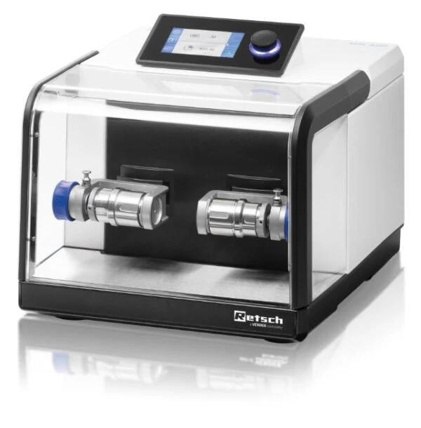
How was periprosthetic tissue prepared in the MM 400 study?
Roux and colleagues treated tissue in sterile single-use bottles with liquid and glass beads at 30 Hz for 210 seconds. The preparation released material for subsequent microbiological examination. Diagnostic interpretation belongs to the laboratory’s complete validated method.
What was processed in the MM 400?
In a 2010 investigation, A.-L. Roux and colleagues used bead treatment to release material from periprosthetic tissue before microbiological examination. They placed the specimen in a sterile 30 mL single-use bottle with 20 mL of sterile water and 5 mL of glass beads measuring 1 mm in diameter.
The reported treatment was 210 seconds at 30 Hz. These figures describe the specimen preparation in that experiment. Recording vessel, liquid and bead volumes alongside milling frequency and duration makes the physical treatment interpretable.
Where did preparation end?
The liquid above the treated specimen was taken forward for culture. Recovery depends on the entire collection, handling and analytical workflow. A frequency and treatment time alone cannot establish diagnostic sensitivity or validate clinical use.
Which specimen and vessel constraints should guide tissue preparation?
An equipment discussion should cover specimen quantity, vessel compatibility and the required preparation endpoint. Diagnostic performance belongs to the complete validated clinical laboratory method. Historical research results do not establish sensitivity or contamination performance for another sample collection or workflow.
| Consideration | Information to provide |
|---|---|
| Specimen format | Tissue quantity and liquid phase |
| Vessel handling | Compatibility and containment |
| Validation | Laboratory controls and analytical endpoint |
Discuss material testing with Glen Mills or Contact Glen Mills for information or a quote.